STAYIN’ ALIVE
a Live Cell imaging workshop at VBCF Advanced Microscopy Facility
12 January 2018
Want to see any of microscopes below in action?
Register before the 9th January 2017 for free demonstrations.
Demonstrations will all take place at the VBCF Advanced Microscopy Facility Labs in Dr. Bohr -gasse 9, Ground Floor, Room 1.005.
Free coupons for lunch at the IMP cafeteria (Campus-Vienna-Biocenter 1) available for the first 45 registered participants!
Be sure to pick up your registration material on the 12th January between 9-10am in front of the VBCF Advanced Microscopy Facility Offices (Dr. Bohr–Gasse 9, Ground Floor, Room 1.005).
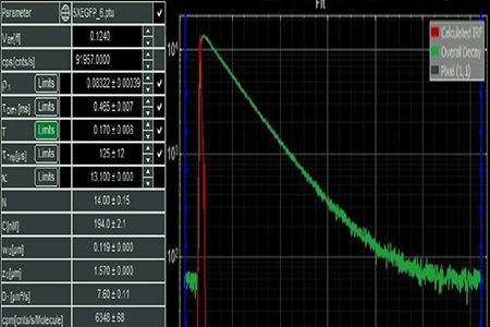
Fluorescence-lifetime imaging microscopy (FLIM)…for measuring fast fluorescence dynamics:The FLIM microscope is a confocal microscope that allows one to, among other things:
|
 |
|
|
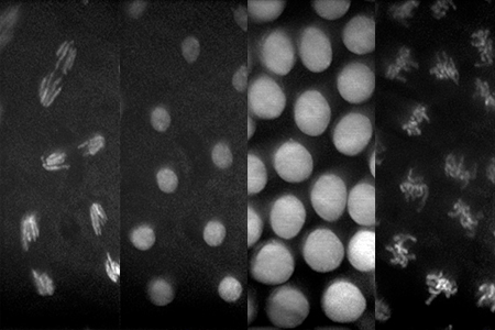
Lattice light-sheet microscope (LLS) …for fast 3D high resolution imaging of sub-cellular and cellular processes:The LLS is a novel light sheet microscope first developed by 2014 Nobel laureate Eric Betzig. It is capable of imaging intra-cellular processes with very high spatio-temporal resolution and minimal phototoxicity, allowing one to study the fast dynamics of life’s fundamental processes in 3D. |
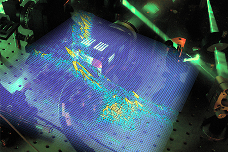
Brillouin Light Scattering Microscope (BLSM)…for ultra-high resolution spectral imaging of a sample to deduce mechanical and structural properties:The BLSM is a microspectroscopy setup that allows for the 3-dimensional label-free mapping of the viscoelastic properties within live cells and tissue, with near diffraction limited resolution. |
 |
|
|
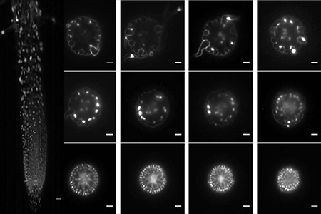
Single Plane Illumination Microscope (LSPIM)…for fast 3D high resolution imaging deep inside tissue:The L-SPIM is a light sheet microscope optimised for high resolution imaging of plant root growth and high-speed volume imaging of neural activity in C. elegans. |